DOI: https://doi.org/10.2497/jjspm.54.75
Translator's note: Author names are written western style, given name first and family name second.
Received November 6, 2006
The microstructure and formation process of hidasuki, a characteristic reddish pattern on traditional Japanese unglazed stoneware called Bizen, was studied through model experiments. Pellets of Bizen clay mined at Bizen-shi/Okayama pref. were heated to 1250C with and without rice straw and then cooled at different rates. A reddish color pattern appeared for relatively slowly cooled samples when rice straw was present. Owing to the presence of potassium in the rice straw, mullite (3(Al,Fe)2O3·2SiO2), a major phase formed in the absence of rice straw, was replaced by corundum (α-Al2O3), hematite (α-Fe2O3), and others in the surface region of about 50μm in depth. The corundum precipitated as hexagonal plate-like crystals, and on the edges of these crystals the hematite grew epitaxially. The growth continued so that the primary corundum crystals were wholly covered by the hematite to form a specific single crystalline α-Fe2O3/α-Al2O3/α-Fe2O3 structure.
Bizen Stoneware, hidasuki, corundum, hematite, epitaxial growth
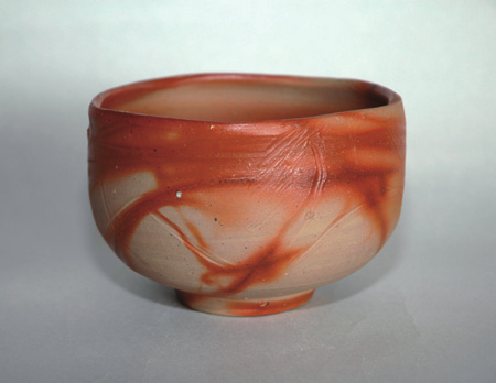
Fig.1 Bizen hidasuki bowl made by Teru Okada.
Bizen ware is one type from among the Six Ancient Kiln sites (Shiragaki, Tokoname, Seto, Echizen, Tamba, and Bizen). Developed from Sue pottery from the Kofun period, it is a traditional pottery with a thousand year long history.
Bizen is called unglazed ceramic; it is complete after a single firing without being glazed. Because the surface is decorated with various colors and patterns, it is known as "art of earth and fire" and its simple elegance is valued in tea ceremonies.
The Bizen patterns include goma, a yellow color created by the reaction between Japanese red pine, used as fuel, and the bare clay; yōhen, from when pottery is buried in ash and charcoal; hidasuki, a red color; and aobizen, which occurs in strong reduction firing. Fig. 1 shows the halmark red color of the hidasuki pattern. The pattern is named after hi [scarlet] colored tasuki [kimono chord], but because it is reminiscent of flames, hi can also be written with the Chinese character for fire.
Bizen ware is unglazed, so it can be packed and stacked during firing, but then pieces may fuse to each other and to the kiln shelves. Rice straw is used to prevent this. When the firing temperature approaches 1200°C, the rice straw leaves the red hidasuki pattern on areas of contact. After the organics in rice straw are burned away, the resulting ash is mainly composed of crystabolite (SiO2), which prevents fusion. It's well known that the red color of hidasuki comes from hematite (iron oxide, α-Fe2O3), the same chemical used for the distinctive red decoration on Kakiemon pottery 1-3). However, the nanostructure and specifics of how hematite precipitates has not been well understood. In this experiment, we sought to understand the nanostructure and formation of hidasuki with transmitting electron microscope analysis 4). We will discuss our discoveries and the unique method by which hidasuki patterns are formed.
Clay from the Kan'on region of Bizen city was mined, levigated, dried, pulverized, mixed, and sieved for particles under 106 μm. The resulting starting material was pressure formed into pellets 20 mm in diameter and about 2 mm thick. Rice straw was placed on top and the samples were heated from room temperature to 1250°C at 1°C/min, then cooled in various ways down to 800°C.
Translator's notes:
A U.S. standard #140 mesh sieve has openings of size 105 μm.
A 1°C/min ramp with peak temperature of 1250°C is equivalent to Orton cone 8.
The test samples were powderized and identified with X-ray diffraction (XRD). The sample surface microformations were imaged with transmitting electron microscope (TEM). The TEM samples were soaked in 1 molar HF solution for 5 minutes, non-glass products that formed within the glass were dispersed with ultrasound and CCl4, and samples were dripped onto a microgrid.
There is a large amount of iron in Bizen clay: if converted to Fe2O3, it represents slightly less than 3wt%. On the other hand, rice straw is high in silicon and potassium. After being heated to 1000°C, the resulting ash is 13wt% potassium oxide (K2O). Hidasuki is formed by the reaction of Bizen clay and the potassium in the rice straw.
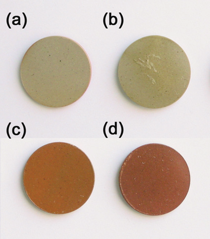
Fig.2 Colors of samples heated with and without rice straw on the surface in air at 1250°C. Sample (a) was heated without rice straw and then cooled to 800°C at a rate of 1°C/min. Samples (b-d) were heated with rice straw and then (b) quenched, (c) cooled to 800°C at a rate of 10°C/min, and (d) cooled to 800°C at 1°C/min. The tone of the reddish color became deeper with decreasing cooling rate.
Fig. 2 shows the surfaces of the Bizen clay with rice straw heated to 1250°C and cooled at various speeds to 800°C. Sample (a) was heated without straw and cooled at a rate of 1C/min. When there is no rice straw, the surface is a yellow-brown color and has a rough texture. There was no change in color regardless of the cooling speed. Sample (b) was heated with rice straw applied and then quenched. The surface was covered in a lustrous glass but there was no red color. Samples (c) and (d) were cooled at 10°C/min and 1°C/min respectively. We show that the slower the cooling is, the deeper the red color becomes. This experiment shows that hidasuki patterns are formed during the cooling after the firing.
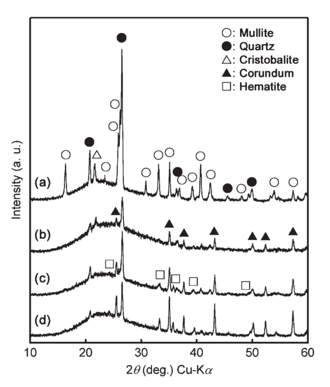
Fig.3 XRD patterns of the surfaces of the samples heated with and without rice straw on the surface in air at 1250°C. Sample (a) was heated without rice straw and tehn cooled to 800°C at a rate of 1C/min (a). Samples (b-d) were heated with rice straw and then (b) quenched, (c) cooled to 800°C at a rate of 10°C/min, and (d) cooled to 800°C at 1°C/min.
The XRD pattern analysis of the sample surfaces in Fig. 2 are shown in Fig. 3. On the untreated sample (a) were quartz (SiO2); crystabolite (SiO2), formed from crystallization of amorphous silica; and mullite (3(Al,Fe)2O3·2SiO2), which forms the ceramic "skeleton". Mullite was not detected on sample (b), which was quenched. Instead there was corundum (alumina oxide, α-Al2O3) and quartz (SiO2). As the cooling rate slowed, the corundum diffraction increased and diffraction appeared and increased for hematite, which is responsible for the red color.This finding is consistent with the colors in Fig 2. Additionally, we find that the samples cooled at 10°C/min and 1°C/min did not develop crystabolite.
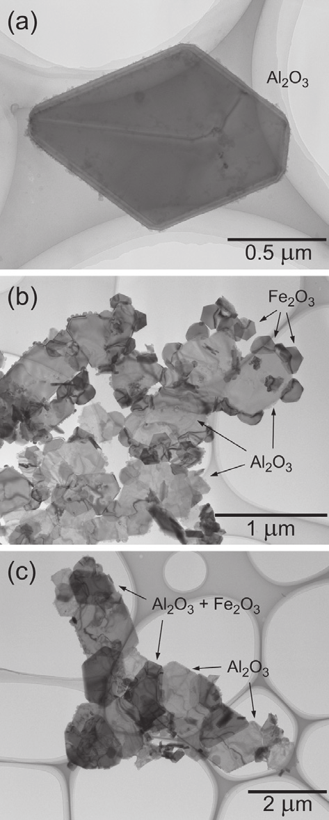
Fig.4 TEM images of crystals in the samples heated with rice straw on the surface in air at 1250°C and then quenched (a) and cooled to 800°C at a rate of 10°C/min (b) and 1°C/min (c). The corundum crystals were formed in the quenced sample (a). The small relatively dark crystals surrounding the corundum crystals seen in (b) were hematite, which grew on crundum epitaxially. In (c) the dark particles marked Al2O3+Fe2O3 are corundum particles wholly covered by hematite like α-Fe2O3/α-Al2O3/α-Fe2O3.
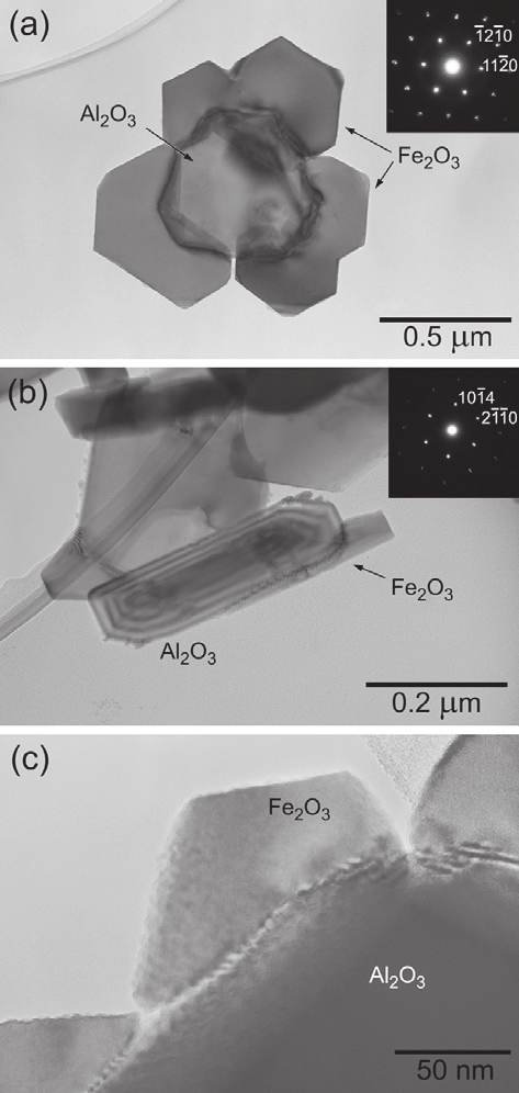
Fig.5 Detailed TEM images and ED patterns of the sample heated with rice straw at 1250°C in air and then cooled at 10°C/min. Image (a) is the [0001] zone axis of corundum and hematite, indicating that hematite epitaxially grew on corundum particles. Image (b) shows tilted image, which indicated that hematite grew from the edges to c-plane of corundum. Image (c) is the boundary region between hematite and corundum.
Fig. 4 shows the pelets that were treated with rice straw, fired, and cooled at various rates the glass that formed on them and their contents scanned by transmitting electron microscope (TEM). It is not displayed on the figure, but the bare clay was composed mainly of the needle like crystals of mullite. The quenched sample in image (a) was composed mainly of the plate shaped corudum. Corundum normally forms grain shaped particles but has been known to form plate shaped particles in the presence of Si, Ca, Mg, and Na ions 6-9). We think plate shaped particles formed because the clay used for this experiment contains all of these elements. (b) is the TEM image of the sample that was cooled at 10°C/min. Attached to the corundum crystals (labeled Al2O3) are small crystals approximately 0.3 um large. From chemical and electron diffraction (ED) analysis, the crystals were identified as hematite. When the cooling rate is further slowed in (c), the corundum crystals grow to 2 um and the hematite crystals completely envelop the larger corundum crystals (labeled Al2O3+Fe2O3). Along with the growth of the hematite, the sample surface becomes redder. Fig. 5 shows the detailed TEM observations of the sample that was cooled at 10°C/min. (a) shows the TEM image of the relatively small corundum particle enveloped by hematite. The ED inset shows the c plane. Since the crystals are completely aligned, it shows that the hematite grows epitaxially on the corundum. Additionally, the fact that morié patterns are observed near the center where the two crystals overlap and the observed intervals (4.4 nm) are consistent with the calculated planar morié intervals confirms the growth type is epitaxial. (b) is the side [0441] image. The hematite grows from the edges of the corundum along the c plane. (c) is the image of the border of the corundum and hematite. The corundum is exceptionally smooth in the c plane but there are some kinks and steps at the edges. These are thought to be the nucleation and growth sites of the hematite. Based on the above TEM observations, we summarize the research results in the schematic in Fig. 6. The reaction between the rice straw and Bizen clay creates a liquid at 1200°C. In this liquid, corundum crystals precipitate and grow as the temperature increases. Depending on cooling conditions, hematite nucleates and grows on the edges of the corundum crystals. Finally, a nucleus of corundum is enveloped in hematite in a core-shell configuration. This is believed to be the cause of the red color.
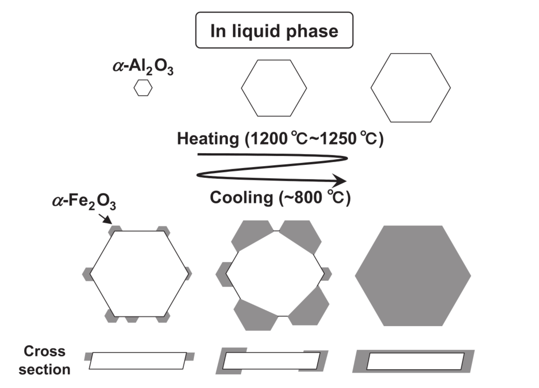
Fig.6 Diagram of the formation mechanism of a core-shell structural particle.
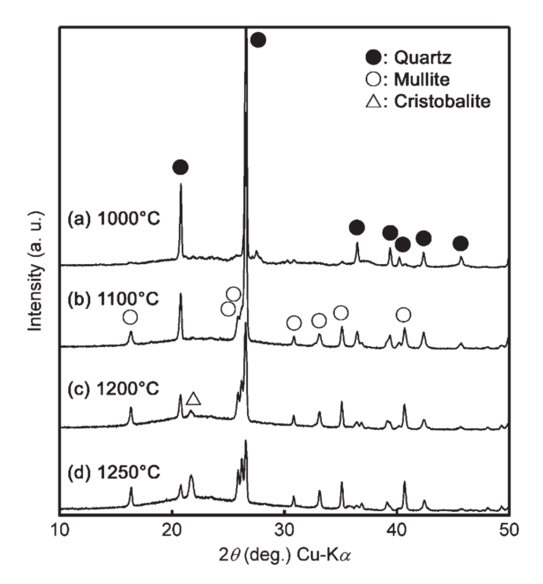
Fig.7 XRD patterns of the Bizen clay heated in air at 1000°C to 1250°C. Mullite formed at 1100°C (b) and cristobalite crystalized at 1200°C (c).
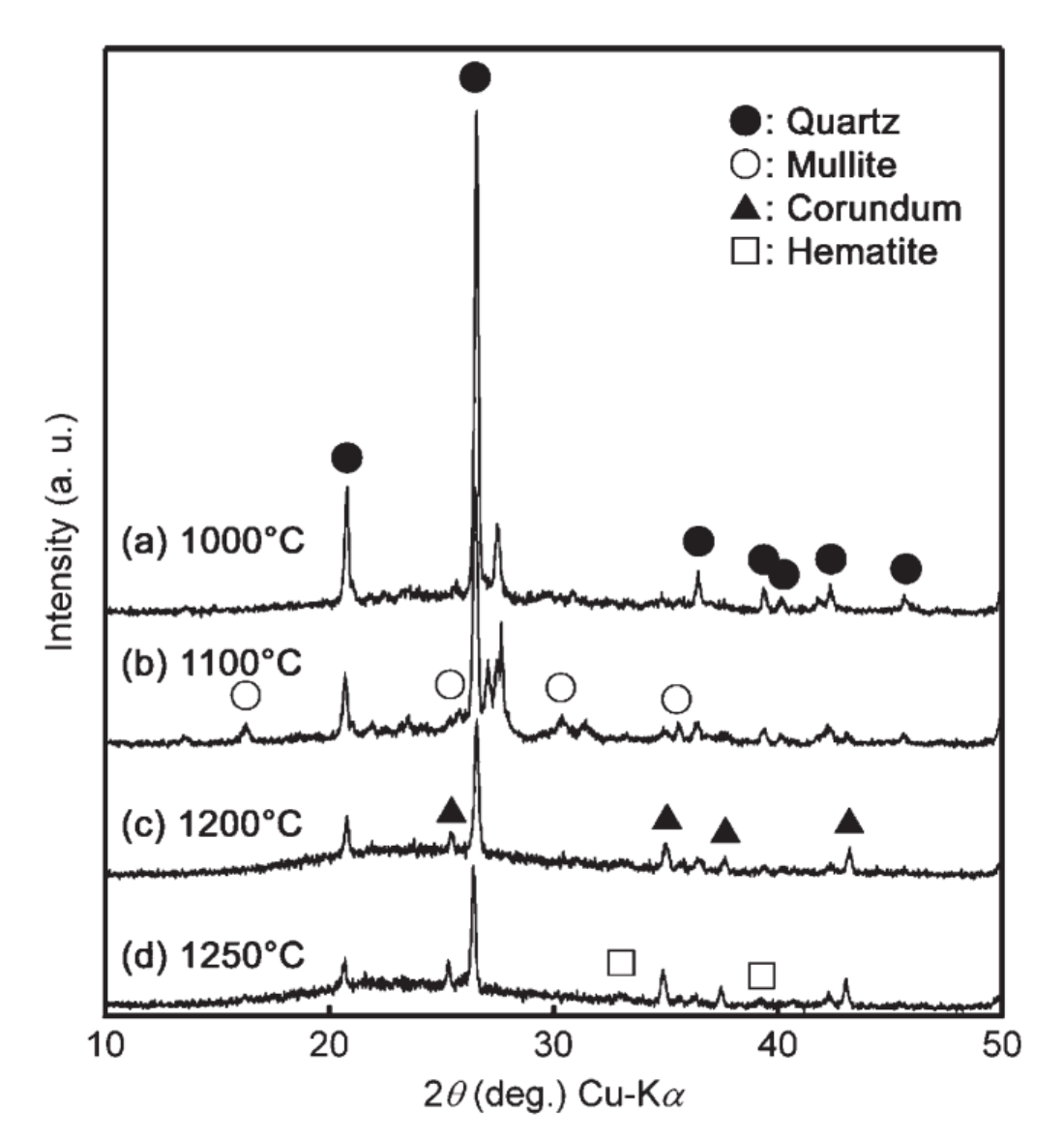
Fig.8 XRD patterns of the samples heated with rice straw in air at 1000°C to 1250°C. Mullite and corundum formed at 1100°C (b) but mullite disappeared at 1200°C (c) completely.
The corundum that forms as the nucleus of the hematite crystals is the key to the red color development of hidasuki. The fundamental oxides that comprise Bizen pottery K2O - Al2O3 - SiO2 are K2O : Al2O3 : SiO2 = 3.1 : 24.6 : 72.3 (wt%) when described in a ternary system. At the triple point of liquid and mullite, corundum can not exist. Even when potassium from rice straw is added corundum can not exist. When bare Bizen clay is heated, it follows the above phase equilibrium. Fig. 7 shows the XRD pattern of the surface of bare Bizen clay after heating and cooling. At 1100°C mullite occurs and at 1200°C crystabolite crystallizes. The halos around 2θ=22° are thought to be from glass phase occuring within the liquid phase. As the temperature increases, mullite formation increases and quartz decreases. Fig. 8 shows the XRD pattern of the surface of the Bizen clay when fired with rice straw. At 1100°C, mullite forms just as in the sample without straw (Fig. 7 (b)), but the quantity is smaller. At this temperature corundum also forms. Above 1200°C, mullite completely disappears.
Therefore, we cannot deny the possibility that mullite growth is suppressed. Mullite is supposed to form at 1100°C, but since there is potassium and iron ions in the melt from the Bizen clay heated in contact with rice straw, it decomposes and corundum forms instead. From this experiment, we discover that when mullite is heated with potassium salt and hematite, mullite is easily dissolved in the corundum and glass phase10).
It is thus shown that plate shaped corundum particles that form from the decomposition of mullite is critical for the formation of hidasuki.
After observation of the microscopic structure and formation process of Bizen Hidasuki pattern, we draw the following conclusions.
The brightness of the red color is greatly hindered by the growth growth of and decrease in distribution of the hematite11-14). However in the case of hidasuki, because it covers the corundum that precipitates earlier during cooling, hematite does not grow and the distribution is preserved. The brightness is preserved even when heated above 1200°C.
This research is made possible by the contributions of Professor Takada and Associate Professor Fujii of Okayama University Grad School; Professor Takano and Adjunct Ikeda of Kyoto University Chemistry Department; Dr. Murakami of Nara Cultural Foundation Research; and Associate Professor Anthony Laurence of Waseda University Engineering Department. To these people we would like to once again express our gratitude. This research was partially funded by the Wakate (B) Research Grant (No. 17750199).
Created .
Home > Q Science > QD Chemistry > Scientific Study of "Hidasuki" Pattern on Bizen Stoneware